An ultrasound machine, also known as sonography equipment, is one of the safest and most commonly used diagnostic imaging tools in modern medicine. From monitoring pregnancy to examining internal organs, ultrasound scans help doctors visualize the inside of the human body without radiation or surgery. Despite its widespread use, many people still ask how an ultrasound machine works and what exactly happens during a sonography test.
This article explains the working principle of an ultrasound machine in a clear, simple, and scientifically accurate way—suitable for patients, students, and anyone interested in medical technology.
What Is an Ultrasound Machine?
An ultrasound machine is a medical imaging device that uses high-frequency sound waves to create real-time images of organs, tissues, blood flow, and developing fetuses inside the body. Unlike X-rays or CT scans, ultrasound does not use ionizing radiation, making it extremely safe.
Ultrasound is widely used to:
- 🤰 Monitor pregnancy and fetal growth
- ❤️ Examine heart function (echocardiography)
- 🫁 Study abdominal organs like liver and kidne
- 🩸 Observe blood flow in vessels (Doppler ultrasound)
- 🦠 Detect cysts, stones, and tumors
Basic Working Principle of Ultrasound
The working principle of ultrasound is based on sound wave reflection (echo).
- The ultrasound machine sends high-frequency sound waves into the body
- These sound waves bounce back (echo) when they hit tissues or organs
- The returning echoes are captured and converted into images
Different tissues reflect sound waves differently, allowing image formation.
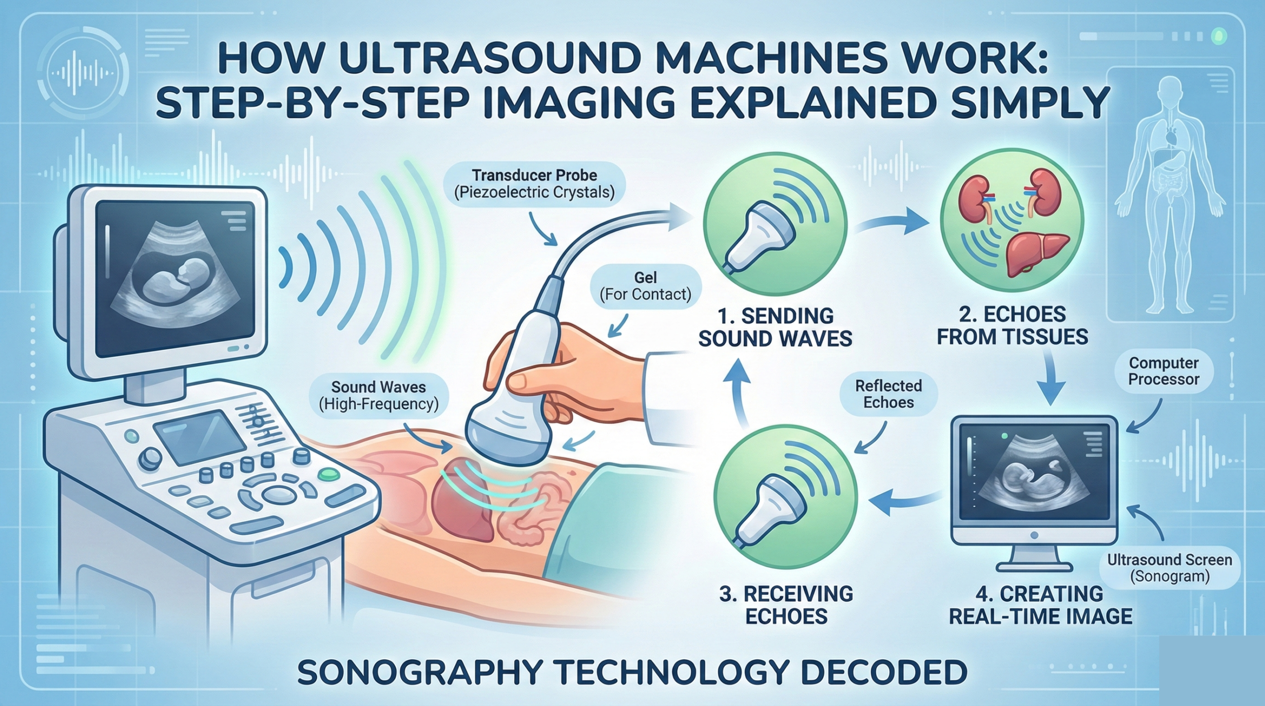
How Ultrasound Machine Works – Step-by-Step Process
Step 1: Generation of Ultrasound Waves
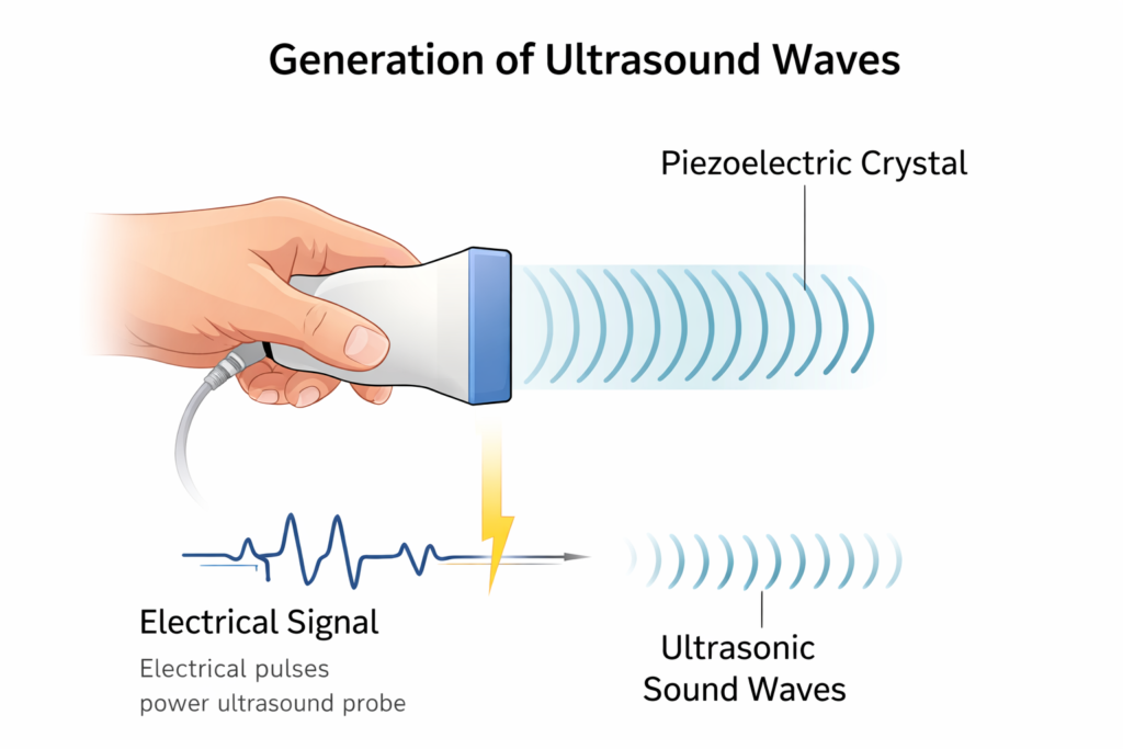
The ultrasound machine uses a device called a transducer (probe).
- The transducer contains piezoelectric crystals
- When electrical current is applied, these crystals vibrate
- This vibration produces high-frequency sound waves
These sound waves are beyond the range of human hearing.
Step 2: Application of Ultrasound Gel
Before scanning:
- A water-based gel is applied to the skin
- The gel removes air between the probe and skin
- Air blocks sound waves, so gel ensures smooth transmission
This step is essential for clear imaging.
Step 3: Sound Waves Travel Through the Body
- The transducer sends sound waves into the body
- These waves travel through tissues at different speeds
- When they hit boundaries between tissues, echoes are produced
Each tissue reflects sound differently.
Step 4: Echo Reception by Transducer
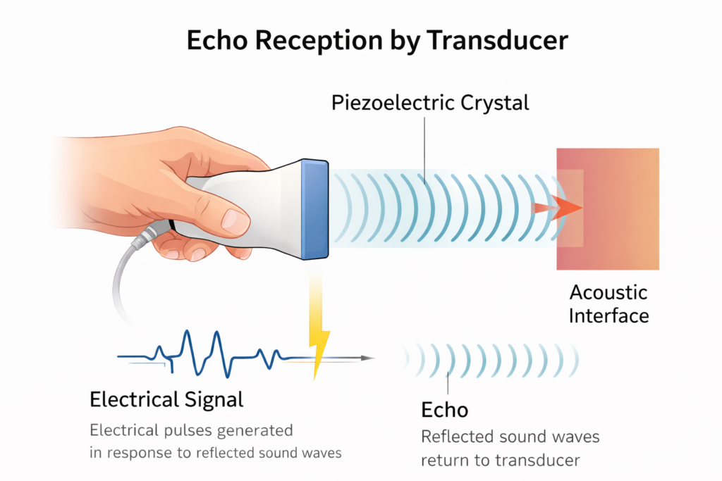
- The same transducer receives the returning echoes
- Piezoelectric crystals convert echoes into electrical signals
- Strong echoes come from dense tissues, weaker ones from soft tissues
This echo pattern forms the basis of the image.
Step 5: Image Formation by Computer

- Electrical signals are sent to the computer system
- The computer calculates distance using echo time
- A real-time image is displayed on the monitor
Doctors can see movement, such as heartbeat or fetal motion.
Main Parts of an Ultrasound Machine and Their Functions

Transducer (Probe)
Generates and receives ultrasound waves.
Computer System
Processes signals and converts them into images.
Display Monitor
Shows real-time ultrasound images.
Ultrasound Gel
Eliminates air gaps and improves sound transmission.
Control Panel
Allows adjustment of frequency, depth, and focus.
Why Ultrasound Images Are Clear and Real-Time
Ultrasound imaging is highly effective because:
- Sound waves reflect differently from tissues
- Real-time imaging shows movement instantly
- No radiation allows repeated scanning
- Doppler technology measures blood flow
This makes ultrasound ideal for dynamic organs like the heart.
Is Ultrasound Scan Safe?
Yes, ultrasound is considered one of the safest imaging techniques.
Safety Advantages
- No radiation exposure
- Non-invasive and painless
- Safe for pregnant women and babies
- Can be repeated multiple times
Unlike X-rays or CT scans, ultrasound has no known harmful effects when used properly.
How Long Does an Ultrasound Scan Take?
- Usually 15–30 minutes
- Complex scans may take longer
- Results are often available immediately
Patients may be asked to change position during the scan.
Ultrasound vs X-Ray vs MRI
| Radiation | ❌ No | ✅ Yes | ❌ No |
| Real-Time Imaging | ⭐ Yes | ❌ No | ❌ No |
| Soft Tissue Imaging | ⭐ Good | ❌ Poor | ⭐ Excellent |
| Pregnancy Safety | ⭐ Excellent | ❌ Not Safe | ⚠️ Limite |
Understanding how an ultrasound machine works helps patients appreciate one of the safest and most versatile diagnostic tools in healthcare. By using high-frequency sound waves, piezoelectric technology, and advanced computer processing, ultrasound machines provide clear, real-time images without exposing patients to radiation. This makes sonography an essential and trusted imaging method in modern medicine.
Read More: